
how AI predicts myopia progression or the technology behind bionic eyes?

AI Predicts Myopia Progression, not by “reading a crystal ball,” but by analyzing massive amounts of eye data to find hidden patterns.👁️

AI Predicts Myopia: Two Different Tasks Current AI performs very differently on predicting onset versus predicting progression rate :· Predicting Onset (Future Myopia): Performs excellently. AI can precisely capture spatial features in the fundus through ResNet34 and track changes over time with LSTM, achieving AUC above 0.94 and even above 0.98 for high myopia .· Predicting Progression (How Fast It Worsens): Currently a global challenge. Because myopia progression is nonlinear, past progression history has limited predictive value for the future .

AI often excels at predicting “will myopia develop” while struggling with “how much will it increase next year” .💡

How AI “Sees Through” Myopia ProgressionAI doesn’t just look at “diopters” but more at ocular biometric parameters:· Monitoring Axial Length: Myopia progression is essentially axial elongation.

AI can identify previously overlooked key predictors. For example, pupil size (importance score 100) and corneal thickness are more critical than age for predicting axial growth rate .·

Extracting Image Biomarkers:

AI extracts subtle features from fundus photographs and OCT (optical coherence tomography) images that are difficult for the human eye to notice, automatically discovering “image biomarkers” related to progression .🔬

The Core Technology of Bionic EyesBionic eyes (visual prostheses) essentially bypass damaged photoreceptors and directly stimulate remaining neurons with electricity. AI plays the role of “translator,” responsible for converting camera images into electrical signals the brain can understand.·

Image Encoding and “Phosphenes”: The camera captures images, and AI processes them into stimulation patterns for phosphenes (light spots).

AI selectively highlights key information like obstacles, ground, and faces to help users navigate independently.·

Hardware Architecture Mimicking the Retina: Advanced research uses Spiking Neural Networks (SNN) to simulate the hierarchical processing of the retina (photoreceptors → bipolar cells → ganglion cells), making electrical stimulation patterns closer to natural neural signals .·

Closed-Loop Optimization: AI algorithms handle image processing while optimizing stimulation strategies in real-time based on user feedback, helping users recognize objects and navigate more easily.

Example like If you’re interested in a specific technology (such as what the images seen through bionic eyes actually look like), we can continue exploring.
How does machine learning and artificial intelligence role in developing and improving eye sight vision?

Machine learning (ML) and artificial intelligence (AI) are transforming eye care across diagnosis, surgical planning, and even vision restoration. Here is how they are developing and improving eyesight.🔍

Early Detection and Diagnosis AI excels at spotting subtle patterns in eye images that may be missed by the human eye.· Refractive Errors & Myopia: Deep learning models analyze fundus photographs to predict refractive power with remarkable accuracy, aiding early detection in children .

AI can predict myopia onset using factors like age, baseline refraction, and axial length .·

Disease Screening: AI algorithms detect diabetic retinopathy on retinal images with sensitivities up to 96% and specificities up to 98% . They also identify keratoconus by analyzing corneal topography with over 98% specificity .

🛠️ Improving Surgical OutcomesAI is being used to personalize and refine vision correction procedures.· Refractive Surgery Planning: Machine learning models predict the best surgical approach (LASIK, SMILE, etc.) with up to 81-93% accuracy .

AI-developed nomograms also achieve a higher percentage of eyes hitting the target refraction post-surgery compared to traditional methods .·

Cataract Surgery: AI improves the calculation of intraocular lens (IOL) power, which is critical for restoring clear vision after cataract removal, especially for patients who had prior refractive surgery .🔬

Vision Restoration TechnologyFor those with severe vision loss, AI is central to developing visual prostheses (bionic eyes).·

Smart Bionic Eye: Researchers are developing AI-driven retinal implants that don’t just replicate vision but provide task-specific visual augmentations. Deep learning filters information to help users with face recognition, navigation, and object identification in real time .·

Optimizing Implants: AI is used to process images into stimulation patterns that the brain can interpret. Spiking neural models and attention mechanisms help the implants mimic how the human retina processes visual information, improving the fidelity of what the user sees .⚠️

Current LimitationsDespite the promise, challenges remain. AI models can function as “black boxes,” and their performance can drop if they are trained on non-diverse datasets (e.g., mostly East Asian or Caucasian populations), leading to biases . Real-world validation is still limited, and results can vary depending on the specific imaging device used .Would you like to dive deeper into a specific area, such as for example how AI predicts myopia progression or the technology behind bionic eyes?

how to improve eye vision?

While you can’t reverse serious eye conditions like cataracts or glaucoma without medical intervention, you can improve your visual comfort, eye health, and focusing ability with these habits.

1. Follow the 20-20-20 Rule Every 20 minutes, look at something 20 feet away for at least 20 seconds. This relaxes the ciliary muscle inside your eye that gets stuck in “near-focus” mode during screen work.

2. Get the Right Prescription If you squint or get headaches, see an optometrist. An outdated or wrong prescription causes constant eye strain. Consider anti-reflective coating and, if you use screens heavily, blue light filtering lenses (though evidence on blue light is mixed, many find it reduces fatigue).

3. Optimize Your Environment· Lighting: Avoid working in a dark room with a bright screen. Use ambient light and position your screen to avoid glare.· Distance: Keep monitors about an arm’s length away, slightly below eye level.· Humidity: Use a humidifier if your eyes feel dry, and blink consciously during screen use.

4. Lubricate Your EyesDry eyes blur vision. Use preservative-free artificial tears if needed. Stay hydrated. Omega-3 fatty acids (fish oil, flaxseed) may help some people with dry eye.

5. Nutrition for Eye Health· Lutein & Zeaxanthin (leafy greens, eggs): Build macular pigment.· Vitamin A (carrots, sweet potatoes): Prevents night blindness.· Vitamin C & E (citrus, nuts): Antioxidants.· Zinc (beans, meat): Helps retina health.· Omega-3s: Supports tear film and retinal health.

6. Eye Exercises (Marginal but Helpful for Strain)· Near-far focus: Alternate looking at your thumb (6 inches away) and a distant object.· Palming: Rub hands warm, cup over closed eyes, relax for 1 minute.· Figure-eight tracking:

Follow an imaginary figure eight with your eyes. These don’t fix refractive errors but can reduce fatigue.

7. Protect Your Eyes· Wear UV400 sunglasses outdoors.· Wear safety glasses for risky activities.· Don’t rub your eyes hard—it can deform the cornea over time (keratoconus).

8. Lifestyle Factors· Sleep: Your eyes repair themselves during sleep. 7–9 hours is ideal.· Exercise: Aerobic exercise lowers eye pressure and improves blood flow.· Blood sugar: Uncontrolled diabetes damages retinal blood vessels.· Smoking: Strongly linked to macular degeneration and cataracts.9. When to See a DoctorGet a dilated eye exam if you notice:· Sudden floaters or flashes· Loss of peripheral vision· Persistent blur or double vision· Eye pain or rednessWhat won’t work: Eye exercises won’t cure nearsightedness, and there’s no diet that lets you throw away glasses. But the habits above can slow decline, reduce strain, and keep your eyes comfortable.Would you like tips specific to screen work, dry eyes, or a certain condition?

how does lens camera work?

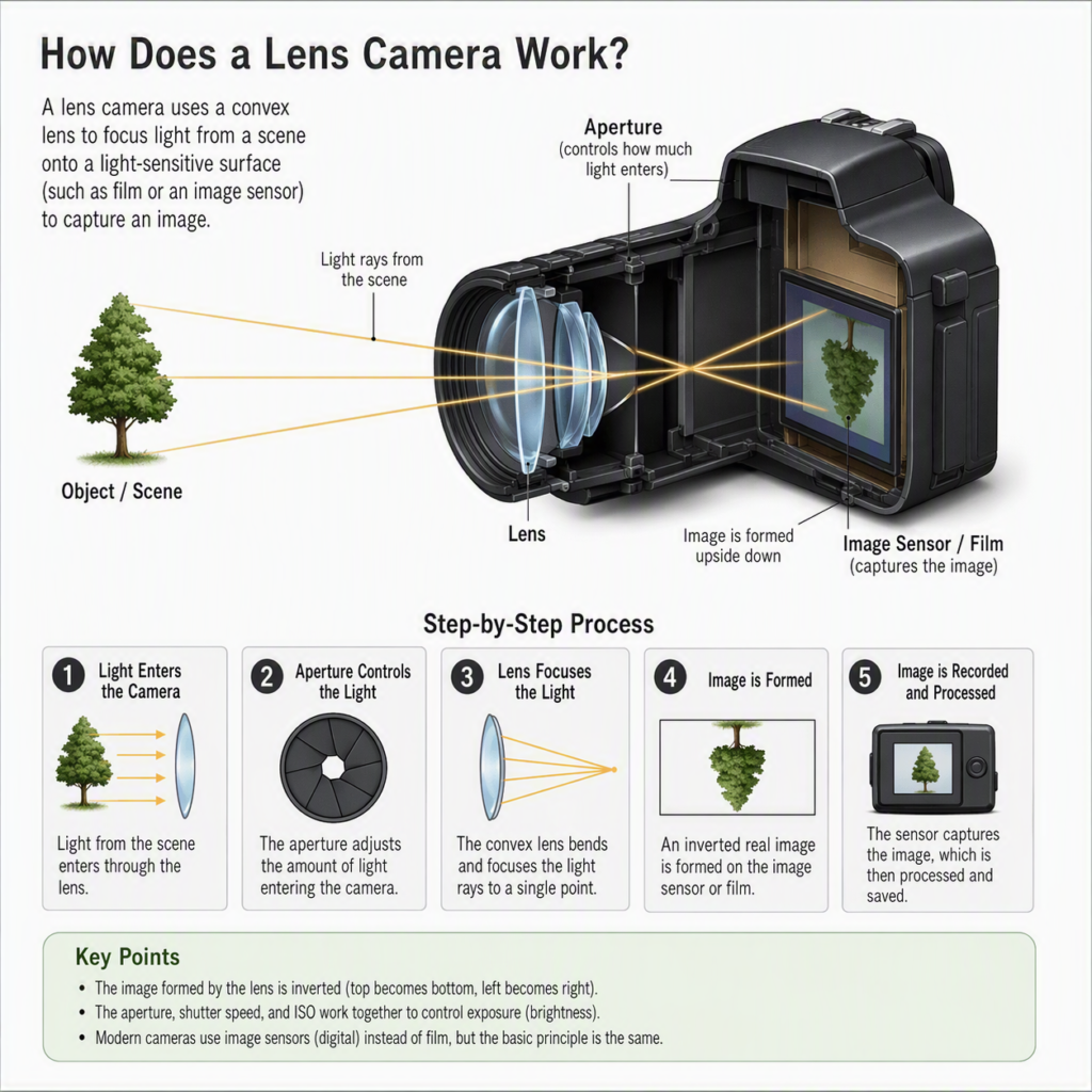
A lens camera works by using a lens to focus light from a scene onto a light-sensitive surface (film or a digital sensor), which records the image.Here’s the basic process:

1. Light enters through the lens Light rays bounce off objects in the scene and travel in many directions. The lens is shaped (usually convex) so it bends these rays.

2. The lens focuses the light The lens bends (refracts) the light rays so they converge and meet at a specific point. This creates a sharp, upside-down image of the scene inside the camera.

3. Focusing adjusts for distance · In a fixed-focus camera, the lens is set so everything beyond a certain distance is acceptably sharp. · In a focusable camera, the lens (or lens elements) moves closer to or farther from the sensor/film so that objects at different distances are in sharp focus.

4. Aperture controls how much light passes The aperture is an adjustable opening in the lens. A larger opening lets in more light (good for dark scenes) but reduces depth of field (less is in focus). A smaller opening lets in less light but increases depth of field.

5. Shutter controls how long light hits the sensor The shutter opens for a set time (shutter speed). Longer times let in more light but risk blur from motion.

6. The image is recorded · Film camera: Light causes a chemical reaction on the film, creating a latent image that’s later developed. · Digital camera: A sensor (CMOS or CCD) converts light into electrical signals, which are processed into a digital image file.7. The image is inverted Because light rays cross at the lens, the image forms upside-down and reversed. The camera (or your brain, when viewing) corrects this.In short: The lens gathers and bends light, the aperture and shutter control the amount and duration of light, and the sensor or film captures the focused image.

how did canon’s lens design philosophy develop

Canon’s lens design philosophy is fundamentally guided by a single core idea: “Enabling photographers to capture the images they envision”. To achieve this, Canon has historically adopted a stance of vertical integration—developing its own materials, manufacturing techniques, and electronics when existing options fell short.🎯

Core Design PillarsCanon’s optical strategy is built on several consistent principles aimed at professional reliability and high performance:·

High Image Quality Across the Frame: Prioritizing sharpness, contrast, and faithful color reproduction from center to edge.·

Natural Bokeh: Designing optical formulas specifically to render smooth, pleasing background blur.· Durability & Reliability: Meeting the rigorous demands of professional use, including weather resistance and robust construction.📜

Key Historical ErasThe philosophy has evolved through distinct technical milestones:· The Fluorite & Aspherical Era (1960s–1970s): Canon pioneered the use of artificial fluorite crystals in 1969 to eliminate chromatic aberration (color fringing) far more effectively than standard glass.

Shortly after, in 1971, they released the FD55mm f/1.2AL, the world’s first SLR lens with a ground aspherical element, which corrected spherical aberration and distortion.· The “L” Series & Autofocus (1978–1980s): The Luxury (L) series was born in 1978, denoted by a red ring, symbolizing the use of these special optical materials. With the 1987 EOS system, Canon shifted to a fully electronic EF mount.

This larger diameter (54mm) was specifically designed to enable ultra-fast f/1.0 lenses and allowed for the Ultrasonic Motor (USM) to be built directly into the lens for silent, fast autofocus.· The Digital & RF Era (2000s–Present): Innovations shifted toward coatings and compact designs.

Canon introduced Subwavelength Structure Coating (SWC) in 2008 to combat ghosting. The 2018 RF mount further leveraged a short flange distance to enable “impossible” optics, such as the RF 28-70mm f/2L, a zoom lens with a constant f/2 aperture that rivals prime lenses.Would you like to dive deeper into a specific technology, such as how Canon manufactures its large fluorite elements?

how are progressive lenses manufactured?

Progressive lenses are manufactured using a combination of mass-produced semi-finished blanks and computer-controlled precision surfacing to create the custom prescription on the back surface.🏭

Mass Production:

Creating the Semi-Finished Blank The complex progressive surface (the corridor) is mass-produced on the front of the lens using cast polymerization:·

Mold Assembly: A glass mold with the progressive curve is paired with another mold and held together by a gasket or tape.

Casting: A liquid plastic monomer (like CR-39) is poured into the cavity and cured in an oven .· Result: This produces a “blank” with a finished front surface but a thick, unfinished back surface .💻

Customization: Free-Form Surfacing To fulfill your specific prescription (including cylinder and prism), the back surface is machined:·

Digital Design: Software calculates the exact 3D shape needed to work with the front progressive curve .·

Precision Machining: A CNC generator mills the back surface, followed by fine turning and polishing .·

Prism Thinning: To keep the lens thin, the back surface is often tilted or cut asymmetrically to distribute thickness more evenly .

🧪 Alternative Methods·

Glass Lenses: Historically made by heating a glass blank until it sags into a progressive mold using vacuum pressure .·

Injection Molding: Used for some materials like polycarbonate, where hot polymer is injected into a precision mold .

Are you interested in the specific materials used, or how the “corridor” design is digitally calculated?

how does lens magnification works with convex lens?

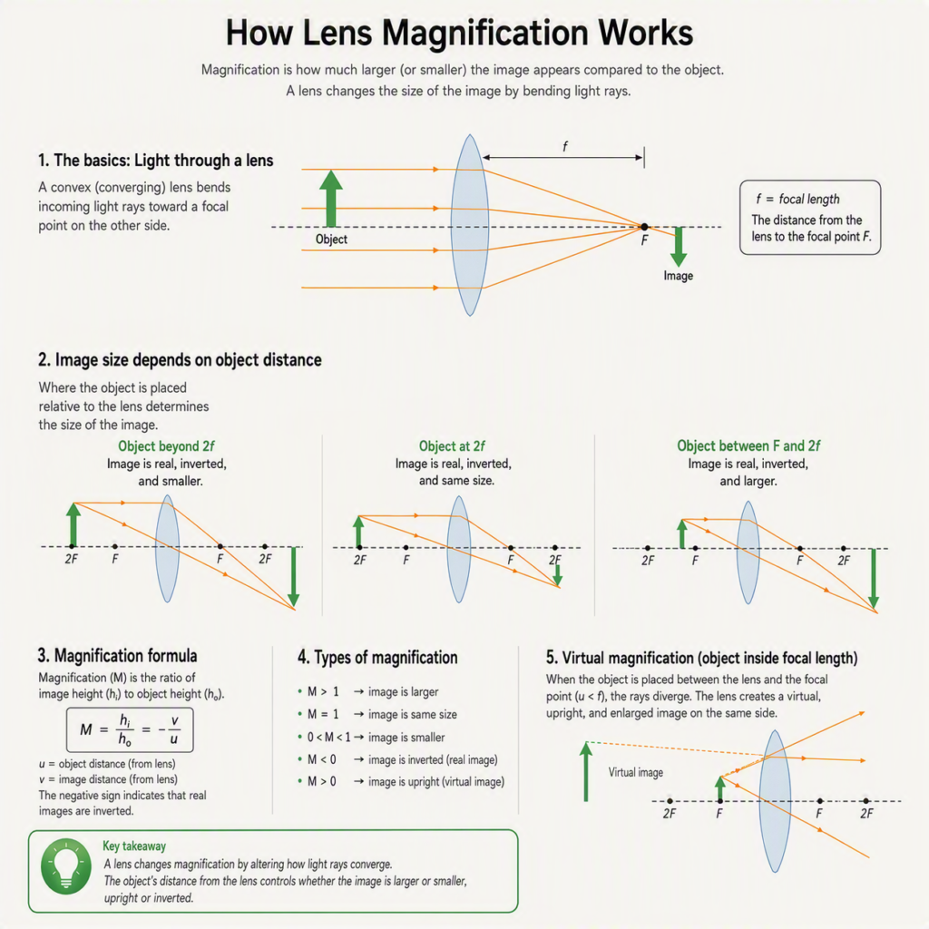
With a convex lens (a lens that bulges outward, thicker in the middle), magnification works through refraction and the geometry of ray diagrams. Here’s how it breaks down:

  1. The Basic Principle: Refraction

A convex lens bends light rays inward (converging them). Parallel rays entering the lens all meet at a single point called the focal point (F). The distance from the lens to this point is the focal length (f).

  1. The Thin Lens Equation

This governs where an image forms:

1/f = 1/dₒ + 1/dᵢ

· f = focal length
· dₒ = object distance (from lens to object)
· dᵢ = image distance (from lens to image)

  1. The Magnification Formula

Magnification (M) tells you how much bigger or smaller the image is compared to the object:

M = -dᵢ / dₒ = hᵢ / hₒ

· hᵢ = image height
· hₒ = object height
· The negative sign means the image is inverted (upside down)

  1. How Magnification Changes with Object Distance

This is the key insight. As you move the object closer to the lens, magnification increases:

Object Position Image Type Magnification
Far away (dₒ > 2f) Real, inverted, smaller M < 1 (reduced) At 2f (dₒ = 2f) Real, inverted, same size M = 1 Between f and 2f Real, inverted, larger M > 1
At f (dₒ = f) No image formed Infinite (rays parallel)
Inside f (dₒ < f) Virtual, upright, larger M > 1 (magnifying glass)

  1. Ray Diagram: How It Actually Works

Three principal rays show where the image forms:

  1. Ray parallel to axis → refracts through the focal point on the other side
  2. Ray through the center → passes straight through undeviated
  3. Ray through the near focal point → emerges parallel to the axis

Where these rays intersect is where the image forms. The farther the intersection is from the axis, the larger the magnification.

  1. The Magnifying Glass Case (Most Common)

When you hold a convex lens closer than f to an object:

· The rays diverge after passing through the lens
· Your eye traces them backward to form a virtual image
· This virtual image is upright and larger
· M = 1 + (25cm / f) for a relaxed eye (25cm = near point of human vision)

Example: A lens with f = 5cm gives M = 1 + 25/5 = 6× magnification

  1. Why Convex Lenses Magnify (Intuition)

Think of it this way:

· A convex lens compresses the angle of incoming light rays
· Your eye receives rays that appear to come from a larger object farther away
· The brain interprets this as a bigger object
· The closer the object is to the focal point, the more extreme this effect becomes

  1. Real vs. Virtual Images

· Real image (dₒ > f): Can be projected on a screen. Inverted. Used in cameras, projectors.
· Virtual image (dₒ < f): Cannot be projected. Upright. Used in magnifying glasses, eyeglasses.

Summary

A convex lens magnifies by refracting light to converge rays, forming an image whose size depends on the ratio of image distance to object distance. Move the object closer to the focal point → magnification increases. Inside the focal point → you get an upright virtual image (magnifying glass). Beyond it → you get an inverted real image (camera/projector).

How does lens magnification work?

Here is a clear breakdown of how lens magnification works, from the basic physics to the practical photography terms.

1. The Fundamental Physics: It’s All About AnglesAt its core, a lens doesn’t actually “magnify” an object like a microscope does. Instead, it changes the angle of view.Imagine looking at a mountain.

With your naked eye, you see a wide scene. If you put a lens in front of your eye, the lens bends (refracts) the light rays coming from the mountain so that they take up a larger portion of your retina. The lens isn’t making the mountain bigger; it’s narrowing the angle of view so the mountain fills more of your vision.

2. The Three Key FactorsMagnification in a lens is determined by three things:· Focal Length (f): This is the distance (in mm) from the lens to the sensor when focused on infinity.

A longer focal length (e.g., 200mm) gives a narrower angle of view and higher magnification. A shorter focal length (e.g., 24mm) gives a wider view and lower magnification.· Distance to Subject (d): The closer you are, the larger the subject appears. Magnification is a ratio of the image size on the sensor to the actual subject size.· Sensor Size: This doesn’t change the lens’s magnification, but it changes the field of view. A smaller sensor (like APS-C) captures only the center of the image circle, making it appear more magnified (this is called crop factor).

3. The Magnification Formula (For Close-Up Work)For macro photography, magnification is expressed as a ratio (e.g., 1:1, 1:2, 2:1).Magnification (M) = Image Size on Sensor / Actual Subject Size· 1:1 (Life Size): A 10mm insect projects a 10mm image on the sensor.· 1:2 (Half Life Size): A 10mm insect projects a 5mm image.· 2:1: A 5mm insect projects a 10mm image (2x magnification).You can calculate this with:M = f / (d – f)(Where f is focal length and d is distance from lens to subject)

4. How Zoom Lenses WorkA zoom lens changes its effective focal length by moving lens elements inside the barrel.· Wide (e.g., 24mm): Light rays are bent gently. The image circle is large, so you see a wide scene.· Telephoto (e.g., 200mm): Light rays are bent more sharply. The image circle is smaller, so you see a narrow, magnified slice of the scene.Important: Zooming does not change perspective. If you stand in the same spot and zoom in, the background appears to compress (larger relative to the foreground), but that’s just because you’re cropping into a smaller area.

5. Magnification vs. ResolutionA common misconception: More magnification ≠ More detail.· If you magnify an image digitally (cropping), you lose resolution.· If you magnify optically (with a longer lens), you preserve resolution because you’re using more of the sensor’s pixels to capture that specific area.· However, every lens has a resolution limit (diffraction). Beyond a certain point (e.g., f/22), magnifying further just makes the image blurrier, not more detailed.6.

Practical Examples· Bird Photography: A 600mm lens on a full-frame camera gives high magnification, filling the frame with a small bird.· Macro Photography: A 100mm macro lens at 1:1 magnification lets you fill the frame with a coin.· Smartphone:

Tiny lenses with short focal lengths. They use digital zoom (cropping) for “magnification,” which degrades quality. Optical zoom on phones uses multiple physical lenses.SummaryLens magnification is the result of focal length narrowing the angle of view, projecting a larger image of the subject onto the sensor.

For close-ups, it’s a ratio of sensor image size to real-life size. Zoom lenses change focal length mechanically, while sensor size affects the field of view, not the true magnification.

What is the use of protective lens?

A protective lens is used to shield the actual lens of a device from damage while maintaining image quality. Its main purposes are:

1.

Physical protection – Guards against scratches, dust, dirt, fingerprints, moisture, and impacts. If it gets damaged, it’s cheaper to replace than the main lens.

2. Preserving resale value – Keeps the original lens in pristine condition, which matters for resale.

3. Optical filtering (in some cases) – Certain protective lenses also add UV filtering, anti-reflective coatings, or glare reduction.

4. Easy cleaning – Can be removed and cleaned without risk of damaging the primary lens coatings.Common uses:· Cameras – Protects expensive camera lenses (often called a “UV filter” or “clear filter”)· Smartphones – Tempered glass or plastic covers over the camera bump· Eyewear/safety glasses – Shields eyes from debris, chemicals, or UV light· Industrial/scientific equipment – Protects sensors, lasers, or optical instrumentsTrade-off: A protective lens can slightly reduce image quality (flare, reduced sharpness) and adds cost, so the choice depends on how valuable the underlying lens is and the shooting conditions.